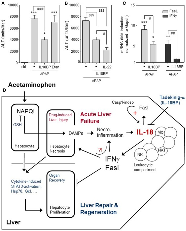
Figure 1.
Effects of IL-18BPd:Fc on APAP-induced ALI. (A–C) All animal experiments (fasted male C57Bl/6 mice, 9–10 weeks old) were carried out in accordance with the recommendations of the Animal Protection Agency of the Federal State of Hessen (Regierungspräsidium Darmstadt, Germany). The protocol was approved by the Regierungspräsidium Darmstadt (Germany). The model of murine APAP (i.p. 500 mg/kg in 0.9% NaCl)-induced liver injury was performed as recently described (46). Where indicated, mice were i.v. cotreated with recombinant IL-18BPd:Fc (IL18BP), IL-22, or etanercept in PBS. (A) Mice received either APAP (n = 18), APAP/IL-18BPd:Fc (15 µg, n = 12), APAP/Etanercept (75 µg, n = 7), or 0.9% NaCl/PBS (ctrl, n = 6). After 24 h, serum alanine aminotransferase (ALT) activity was determined (Reflotron, Roche Diagnostics, Mannheim, Germany) and is depicted as units/liter (means ± SEM). *P < 0.05, ***P < 0.001 compared to ctrl; ###P < 0.001. (B) Mice received either APAP/PBS (n = 12), APAP/IL-18BPd:Fc (15 µg; n = 12), or APAP/IL-18BPd:Fc (15 µg) plus IL-22 (3.5 µg) (n = 13). After 24 h, serum ALT activity was determined and is depicted as units/liter (means ± SEM). $$$P < 0.001, #P < 0.05. (C) Mice received either APAP/PBS (n = 12) or APAP/IL-18BP (15 µg, n = 12) and were maintained for 16 h. For RNA analysis, liver tissue was snap frozen in liquid nitrogen and stored at −80°C. Total RNA was isolated as described (18). For real-time PCR, pre-developed reagents were used (Thermo Fisher Scientific, Darmstadt, Germany): GAPDH (VIC; 4352339E), Fas ligand (FasL) (FAM; Mm00438864_m1), and IFNγ (FAM; Mm01168134_m1). Assay mix was from Nippon Genetics (Düren, Germany). PCR: one initial step at 95°C (2 min) was followed by 40 cycles at 95°C (5 s) and 62°C (30 s). Detection of the dequenched probe, calculation of threshold cycles (CT values), and data analysis were performed by the Sequence Detector software (AbiPrism7500 Fast Sequence Detector, Thermo Fisher Scientific). Relative changes in hepatic FasL [(C), left panel] and IFNγ [(C), right panel] mRNA expression determined by real-time PCR were normalized to that of GAPDH and shown as fold-induction compared with untreated control mice (n = 6). *P < 0.05, **P < 0.01, ***P < 0.001 compared with untreated control; #P < 0.05, ##P < 0.01. (A–C) Data are shown as means ± SEM. Raw data were analyzed by one-way ANOVA with post hoc Bonferroni correction. (D) Graphical summary of processes affecting outcome of APAP-induced ALI with focus on the pathogenic role of IL-18. Detrimental pathways activated by APAP overdosing are counteracted by endogenous mechanisms supporting organ recovery through repair and regeneration [e.g., hepatocyte STAT3 activation; expression of heat shock protein (Hsp)70 and glutamate-cysteine ligase (Gcl)]. If therapeutic NAC intervention aiming at augmentation of hepatocyte glutathione (GSH) fails due a to an exceedingly high APAP dosage, a too late time point of intervention, and/or a pre-damaged liver parenchyma, acute liver failure may proceed to an ill-fated condition requiring transplantation for patient survival. Here, IL-18 may play a unique role by supporting hepatic expression of FasL and IFNγ. Application of recombinant IL-18 binding protein (Tadekinig-a) may evolve as a novel therapeutic option to intervene at this point.

