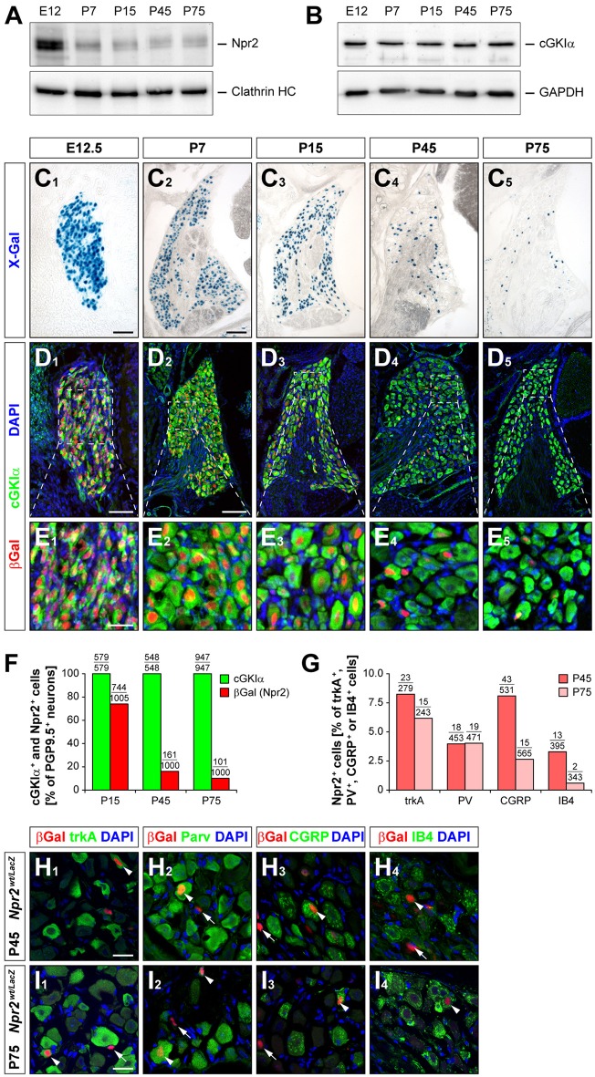
Figure 5.
Expression of Npr2 and cGKIα in DRGs at different developmental stages. (A,B) Western blot of membrane or cytosolic fractions of DRGs using antibodies to Npr2 or to cGKIα, respectively. The heavy chain of clathrin or GAPDH served as loading control. (C–E) Sections through thoracic DRGs at different stages. Localization of anti-βGal staining indicating Npr2-expression is demonstrated in the Npr2wt/LacZ-mouse reporter and localization of cGKIα by a polyclonal antibody to mouse cGKIα. Scale bars in (C1,D1), 50 μm; in (C2-C5,D2-D5), 100 μm; in (E) 25 μm. (F) Quantification of Npr2-positive and cGKIα-positive neurons in DRGs counterstained with an antibody to PGP9.5 at different stages. (G–I) A small proportion of Npr2-positive DRG neurons at P45 and P75 express trkA, parvalbumin, CGRP or IB4. Scale bars in (H,I), 20 μm.

