Fig. 6.
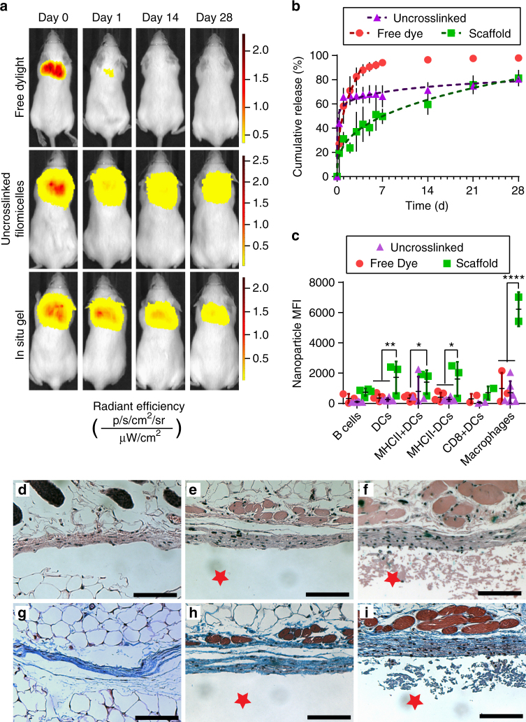
In vivo sustained release from in situ-crosslinked filomicelle scaffolds over one month. a Representative IVIS images of Dylight 755 injections as: free solubilized dye, conjugated to 755-BCP modules within uncrosslinked three-component modular FMs, or conjugated to 755-BCP modules within four-component FMs of in situ crosslinked scaffolds. Radiant efficiency intensity scaled per individual mouse. b Cumulative release curves and power law model fits of MC (Dylight 755) release from FM-scaffolds. c Flow cytometric analysis of MC (Dylight 633) uptake by phagocytic immune cell populations. Significance determined by Tukey multiple comparison test: *p < 0.05, **p < 0.01, ****p < 0.0001. For b and c, error bars represent s.d, n = 5 mice for free dye and n = 4 mice for uncrosslinked and in situ groups. Representative images of H&E staining of the interface between skin and d the saline Dylight 633 solution-injected control; e uncrosslinked FMs; or f the in situ crosslinked FM-scaffold, respectively. g, h, i, Representative images of Masson’s Trichrome staining for the same groups listed above, respectively. The red star represents the scaffold or uncrosslinked FM side of the interface, for orientation purposes. All images shown are at 10× objective magnification, scale bars are 100 µm

