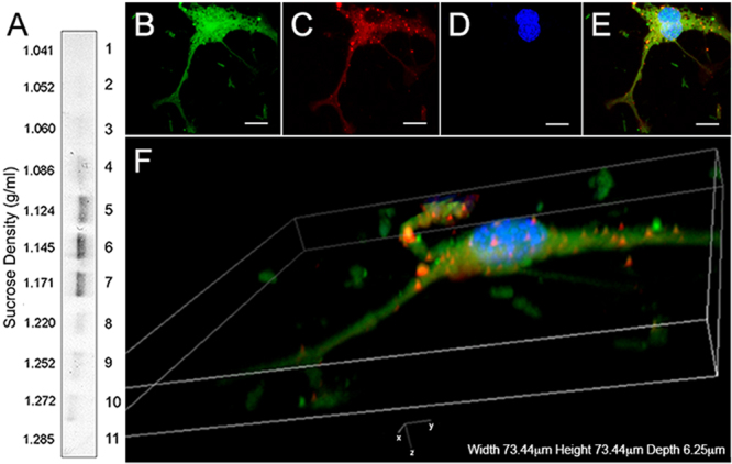
Figure 3.

Sucrose-gradient and immunohistochemical characterization of extracellular vesicles. (A) CD63 positive bands were detected in fractions at gradient-densities of 1.12–1.17 g/cm3. (B–F) Immunohistochemical analysis EVs: (B) GFP+ mRPCs (green), (C) anti-CD63 labeling in cytoplasmic and nuclear regions (red), (D) DAPI (nuclei, blue) and (E) overlay. Scale: 10 µm. (F) 3D confocal reconstruction of labeled mRPCs revealed the presence of CD63 positive EVs in the cytoplasm, emerging from lipid bilayer regions of cell soma and on proximal and distal processes.
