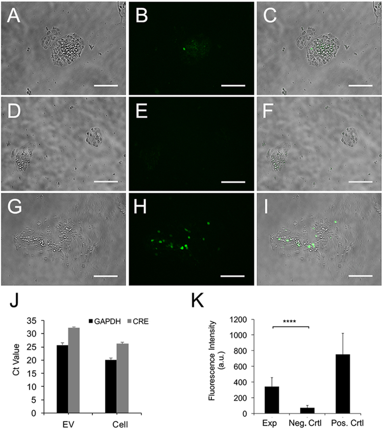
Figure 7.
Extracellular vesicle functional transfer of Cre in dissociated mRPCs. P0 retina were electroporated with Cre+ plasmid, dissociated into mRPCs and plated in 0.4 µm pore transwell inserts. Dissociated mRPCs electroporated with reporter constructs were placed in lower wells beneath Cre+ mRPCs. (A–C) EV mediated functional transfer of Cre leading to activation of GFP expression in reporter RPCs as can be observed in experimental wells following 14 days of culture. (D–F) Negative controls, containing reporter constructs electroporated mRPCs, did not show GFP expression. (G–I) Positive controls, electroporated with both Cre+ and reporter constructs, showed robust GFP expression. Scale: 100 µm. (J) Real time PCR verified Cre+ signal in EV derived from Cre+ electroporated cells. (K) Analysis of GFP intensity in mRPCs revealed a significant difference between experimental and neg. control (experimental: mean 341.41, sd115.92, neg.control mean 70.39 sd 31.65) P value < 0.0001 experimental vs. negative control and between experimental and pos.control (751.42 sd 271.1293979) P value = 0.05 experimental vs positive control.

