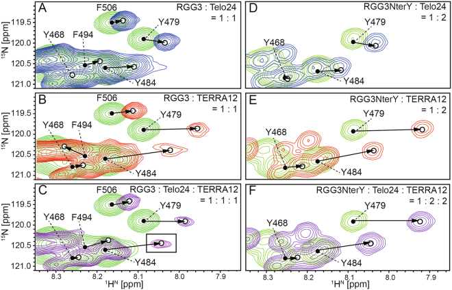
Figure 7.
Chemical shift perturbation patterns for the amide 1H and 15N resonances of tyrosine and phenylalanine residues of either RGG3 or RGG3NterY. An equivalent amount of either Telo24 (A), TERRA12 (B), or both (C) was added to [13C, 15N]-labeled RGG3. Two equivalent amounts of either Telo24 (D), TERRA12 (E), or both (F) were added to [13C, 15N]-labeled RGG3NterY. The spectra of free (green) and complex (either blue, red, or purple) RGG3 are overlaid. The amide resonances of tyrosine and phenylalanine residues of RGG3 in its free form (closed circles), and in either the RGG3:quadruplex binary or ternary complex (open circles), which were both assigned by the triple-resonance NMR method, are connected by arrows. An inset in C is shown with 2-fold lower contour level.

