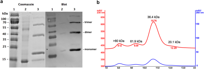
Figure 2.
recMgIFN-γ purified by immobilised metal affinity chromatograph is mostly in a dimeric form. (a) SDS-PAGE followed by Coomassie staining for total protein detection (left) and detection of 6His-tagged proteins by Western Blot with an anti-6His monoclonal antibody (right). Lane 1: molecular weight marker (with sizes given to the left - sizes greater than 100 kDa were unreliable when constructing a standard curve); lane 2: flow-through (unbound proteins); lane 3: purified protein(s). (b) Size determination of purified recMgIFN-γ by analytical gel filtration analysis on a pre-calibrated Superdex 75 column. Protein peaks were detected at 230 nm (red) and 260 nm (blue), respectively.

