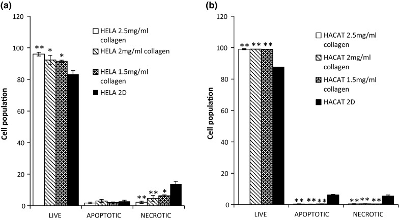
Fig. 4.

YOPRO and PI stained flow cytometry live, apoptotic and necrotic assay for HeLa (a) and HaCaT (b) cells grown on Collagen (3D) in different concentration and cells grown on plastic (2D) culture. Data are expressed as a percentage of three independent experiments ± SD of three individual experiments. Statistically significant differences between the 3D culture membrane live/dead cell analyses and that of the 2D cultures are denoted by *P < 0.05 and **P < 0.01
