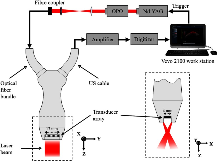
Fig. 1.
PAI system used to scan pigmented skin lesions. (Left) Schematic of PAI system showing laser source, signal processing procedure, and linear-array PA probe (viewed from elevational direction). (Right) View of the PA probe head (enclosed by dashed box on left) showing crossed laser beam geometry (lateral direction).

