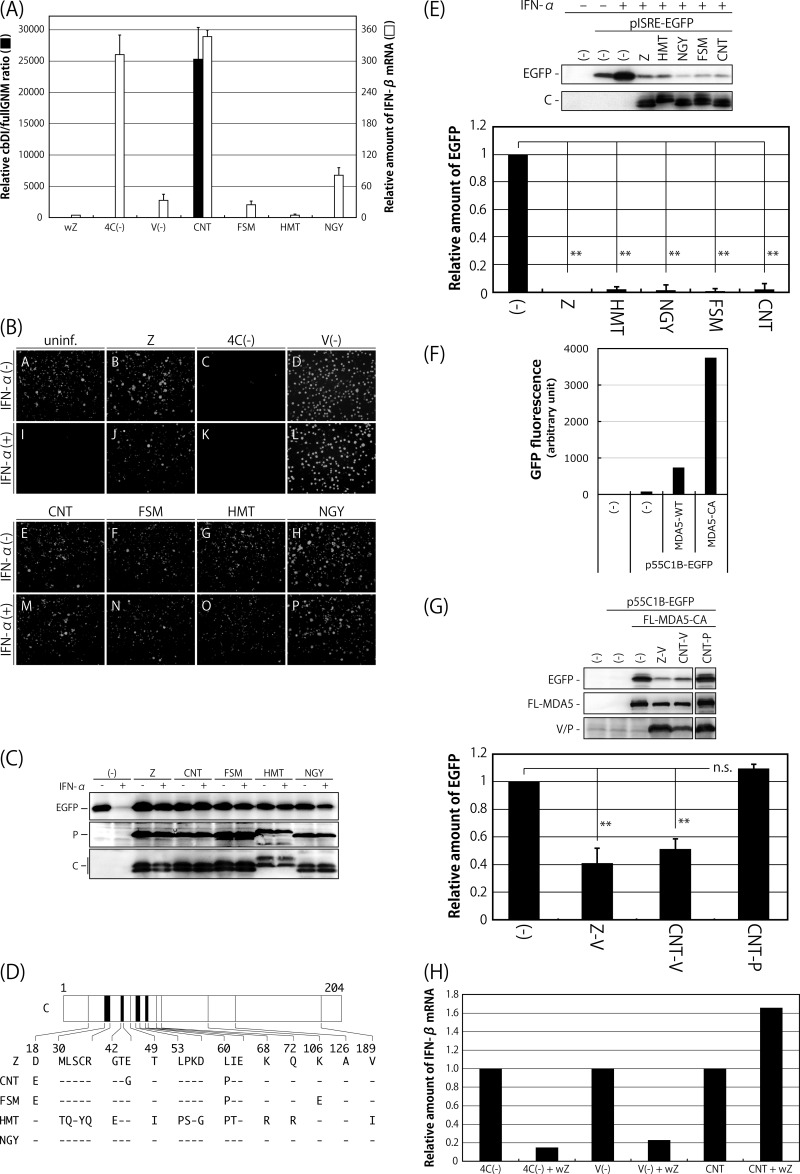
FIG 1.
Comparison of SeV strains in terms of the content of cbDI particles in their working stocks, induction of IFN-β and the antiviral state, and counteraction against the type I IFN pathway. (A) The amounts of cbDI genomes and fullGNM in the working stocks of the indicated viruses were analyzed by one-step qRT-PCR. The cbDI/fullGNM ratios are shown. The ratio of the wZ sample was set to 1. The amounts of IFN-β and beta-actin mRNAs in the infected HeLa cells were also analyzed by one-step qRT-PCR in cells infected with the indicated viruses. The ratios of IFN-β to beta-actin mRNAs are shown. The ratio in the uninfected sample was set to 1. (B) HeLa cells were infected with the indicated viruses. At 6 h p.i., the media were replaced with fresh media containing IFN-α (1,000 IU/ml) or no IFN-α. After an additional 6-h incubation, the cells were superinfected with rVSV-GFP. After further incubation for 6 h, GFP expression derived from rVSV-GFP replication was observed under a fluorescence microscope. (C) Western blotting of the cell lysate samples in panel B using anti-GFP, -SeV P, and -SeV C polyclonal antibodies. (D) Schematic representation of the C proteins of the indicated SeV strains. Dashes indicate that the amino acids are identical to those of Z strain. (E) 293T cells were cotransfected with the C proteins derived from the indicated SeV strains, together with a reporter plasmid, pISRE-EGFP. At 18 h p.t., the cells were treated with IFN-α (1,000 IU/ml) for 8 h, and then the expression level of EGFP was analyzed by Western blotting using an anti-GFP antibody. The amount of EGFP in the cell lysates was quantitated and graphed. The value of mock-transfected and non-IFN-α-treated samples was set to 1. (F) 293T cells were cotransfected with MDA5-WT or -CA, together with a reporter plasmid, p55C1B-EGFP. At 24 h p.t., GFP fluorescence was measured using a fluorometer. (G) 293T cells were cotransfected with the V or P proteins derived from the indicated SeV strains, together with a reporter plasmid, p55C1B-EGFP, and pCAG-FL-MDA5-CA. At 24 h p.t., the expression level of EGFP was analyzed by Western blotting using an anti-GFP antibody. The amounts of EGFP in the cell lysates were quantitated and graphed. The value of the sample receiving FL-MDA5-CA but no V protein was set to 1. (H) HeLa cells were infected with SeV strain Z at an MOI of 5. At 6 h p.i., the cells were superinfected with strain Cantell, Z-4C(−), or Z-V(−). After an additional 24 h of incubation, the amounts of IFN-β and beta-actin mRNAs were analyzed by one-step qRT-PCR. The ratios of IFN-β to beta-actin mRNAs are shown. The ratio in the cells without superinfection was set to 1. All of the bar graphs represent the averages of three independent experiments, and the error bars represent the standard deviations. n.s., nonsignificant (P > 0.05); **, P < 0.01 by one-way analysis of variance (ANOVA) with Bonferroni post hoc test.

