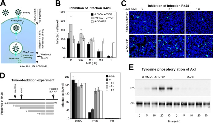
FIG 6.
Virus-induced Axl tyrosine kinase activity is required for rLCMV-LASVGP entry. (A) Schema of the inhibitor washout experiment. For details, please see the text. LE, late endosome. (B) Infection of rLCMV-LASVGP depends on the activity of the Axl tyrosine kinase. HT-1080 cells were pretreated with the Axl tyrosine kinase inhibitor R428 at increasing concentrations for 30 min, followed by infection with the indicated viruses at 200 PFU/well in the presence of drug. After 1 h, cells were washed 3 times with medium containing 20 mM ammonium chloride, followed by 16 h of incubation in the presence of the lysosomotropic agent. Infection was detected by IFA as described for Fig. 3B. Data are means ± SD (n = 3). (C) Example of the inhibition of rLCMV-LASVGP infection by R428 revealed by IFA using MAb 113 to LCMV NP (green) and counterstaining of nuclei (DAPI; blue). Note the similar intensities of NP staining with increasing inhibitor concentrations (bar = 50 μm). The lower panel shows AdV5-GFP used as a negative control. NP- or GFP-positive cells were scored, with cell doublets considered single infectious events. (D) Time-of-addition experiment. HT-1080 cells were infected with rLCMV-LASVGP (300 PFU/well), with R428 or ribavirin (Rib) added at the different time points pre- or postinfection at 0.5 μM or 100 μM, respectively. Virus was added at time point 0, followed by washing and addition of ammonium chloride after 1 h. Ammonium chloride was kept throughout the experiment, and cells were subjected to IFA after 16 h. Data are means ± SD (n = 3). Examination of cell viability by the CellTiter-Glo assay reveled no significant toxicity of R428 under our experimental conditions (data not shown). (E) Engagement of rLCMV-LASVGP induces activation of Axl tyrosine kinase. Serum-starved HT-1080 cells were incubated with rLCMV-LASVGP at 50 PFU/cell and equivalent amounts of mock preparation for 2 h in the cold. Unbound virus was removed, and cells were shifted to 37°C. At the indicated time points, cells were lysed and Axl was isolated by IP with polyclonal Ab goat anti-Axl immobilized on a Sepharose matrix. Immunocomplexes were eluted, followed by SDS-PAGE using 5.5% (wt/vol) running gels. Total Axl protein in IP was revealed in a Western blot using a mouse MAb, anti-Axl (10% of sample), and tyrosine phosphorylation was detected by an MAb to tyrosine phosphate (90% of sample). For detection, TrueBlot HRP-conjugated anti-mouse IgG was used in ECL assays. Blots for tyrosine phosphate (PY) were exposed for 2 min and blots detecting total Axl for 10 s.

