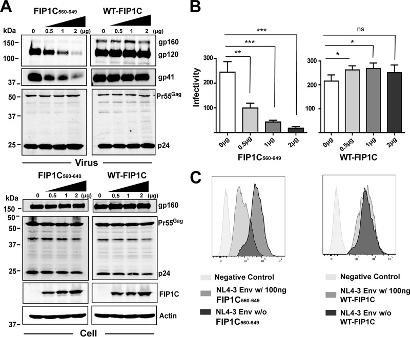
FIG 1.
Truncated FIP1C inhibits Env incorporation and particle infectivity. (A) GFP-FIP1C and truncated mutant of FIP1C (FIP1C560–649) plasmids were transfected in increasing concentrations together with a fixed amount of pNL4-3 proviral DNA into HeLa cells. Shown are results of this titration on viral protein content (upper blots) and cellular protein content (lower blots) 48 h posttransfection. (B) Infectivity of virus particles was quantified using TZM-bl cells following dose titration of FIP1C560–649 (left) or wild-type FIP1C (right). Results are shown as blue cells/nanogram of p24 and are presented as means ± standard deviations (SD). Statistical comparisons between groups were performed using the unpaired t test. Graphs shown are representative of major findings from three independent experiments. ns, ρ > 0.05; *, ρ < 0.05; **, ρ < 0.001; ***, ρ < 0.0001. (C) Cell surface staining for Env using anti-gp120 mouse monoclonal antibody BDI123 (Novus Biologicals) 48 h following transfection with the indicated constructs. Cells first were gated for GFP-FIP1C expression.

