
FIG 1.
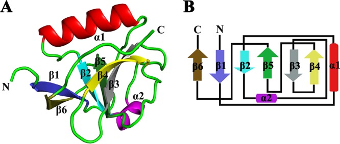
Crystal structure of PEDV nsp1. (A) Crystal structure of monomeric PEDV nsp1. The structure of nsp1 is shown as a cartoon in colors ranging from blue in the N-terminal region to brown in the C-terminal region. (B) Topology diagram. The colors correspond to those in panel A. Strands β1, β2, β3, β4, β5, and β6 make up the barrel.