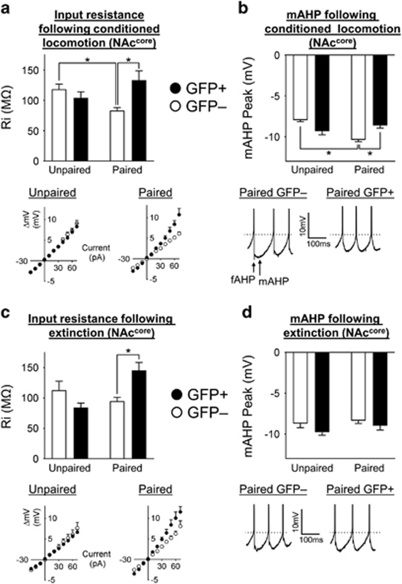
Figure 5.
Modulation of input resistance and AHP underlies excitability changes following cocaine and extinction (EXT) memory retrieval in the nucleus accumbens core (NAccore). (a) The input resistance of GFP+ neurons in Paired conditioned locomotion (CL) mice was increased compared with Paired CL GFP− neurons. Also, the Paired CL GFP– neurons exhibited decreased input resistance compared to the Unpaired CL GFP– neurons.. In Unpaired CL mice, the input resistance of GFP+ and GFP− neurons was similar. Below: I/V curves of Paired CL and Unpaired CL GFP+ and GFP− neurons from which input resistance was calculated. In the Paired CL group, there was a significant shift in the I/V curve of GFP+ compared with GFP− neurons; in contrast, I/V curves of Unpaired CL GFP+ and GFP− neurons were similar. (b) Following cocaine memory retrieval, the mAHP of Paired CL GFP− neurons was significantly increased compared to Paired CL GFP+ and Unpaired CL GFP− neurons. Below: example traces of Paired CL GFP+ and GFP− neurons following cocaine memory retrieval, identifying the position of fast afterhyperpolarization (fAHP) and medium afterhyperpolarization (mAHP) peaks. Dashed line indicates the threshold of the first spike labeled with group means. The fAHP and mAHP of Paired CL GFP− neurons is increased following cocaine memory retrieval. Scale bar, 10 mV and 100 ms. (c) Following EXT memory retrieval, the input resistance of Paired EXT GFP+ neurons was increased compared with Paired EXT GFP− neurons. In the Unpaired EXT mice, the input resistance of GFP+ and GFP− neurons was similar. Below: I/V curves of Paired EXT and Unpaired EXT GFP+ and GFP− neurons from which input resistance was calculated. In the Paired EXT group, the I/V curve of GFP+ neurons was significantly shifted compared with GFP− neurons; in contrast, the I/V curves of Unpaired EXT GFP+ and GFP− neurons were similar. (d) The mAHP of GFP+ and GFP− neurons was not significantly different in either Paired EXT or Unpaired EXT mice. Below: example traces of Paired EXT GFP+ and GFP− neurons. The fAHP and mAHP of Paired EXT GFP+ and Paired EXT GFP− neurons were similar following EXT memory retrieval. Dashed line indicates the threshold of the first spike labeled with group means. Scale bar, 10 mV and 100 ms. All data are expressed as mean±SEM; *p<0.05.

