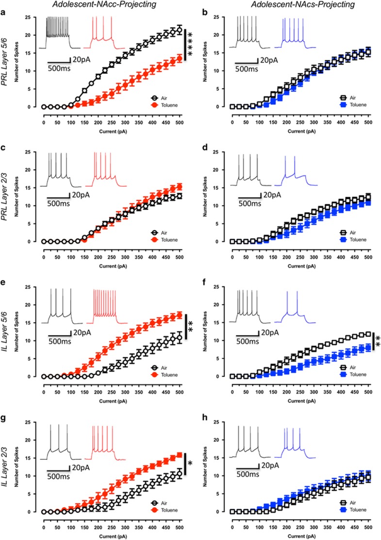
Figure 3.
Toluene exposure produces sub-region and projection-target selective alterations in current-evoked firing of adolescent mPFC neurons. Figures show the effect of toluene exposure on firing of prelimbic and infralimbic mPFC neurons projecting to the core (a—Air: n=11, Toluene: n=12; c—Air: n=7, Toluene: n=7; e—Air: n=8, Toluene: n=9; g—Air: n=6, Toluene: n=7) or shell (b—Air: n=8, Toluene: n=8, d—Air: n=7, Toluene: n=7; f—Air: n=9, Toluene: n=10; h—Air: n=7, Toluene: n=8) of the NAc. Traces in each figure show examples of firing (evoked with a 250 pA current step) from air (left trace) or toluene (right trace) exposed rats measured 24 h after exposure to 10 500 p.p.m. toluene vapor. Data are presented as mean and SEM. Symbol (*) indicates main effect of toluene treatment on firing (two-way repeated measures ANOVA; p<0.05).

