
Fig. 5.
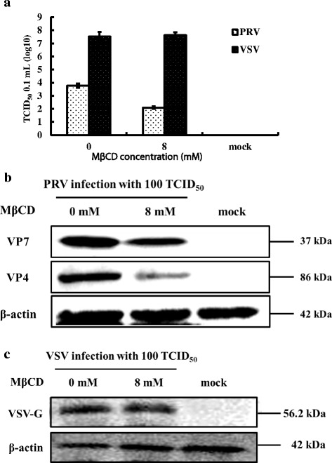
Cholesterol and control of PRV entry. MA104 cells (PRV) and BHK-21 cells (VSV) were either mock treated or treated with 8 mM MβCD for 30 min then infected with PRV or VSV (100 TCID50) for 1 h at 37 °C. Antibodies to the PRV VP7 or VP4 proteins and to the VSV G protein were used to screen western blots of cell homogenates and virus titers. a Virus titers of PRV and VSV after treatment. b Western blot of PRV VP7 and VP4 proteins and β-actin. c Western blot of VSV G protein and β-actin. Western blots depict representative experiments. Error bars (Fig. 5a) indicate the standard deviations of three independent experiments