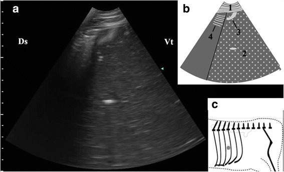
Fig. 4.

Ultrasonogram of interface between abomasal gas and ingesta (a), schematic diagram (b) and position of transducer (c). A 3.5 MHz low-frequency curvilinear transducer was placed in the median part of 10th ICS on the left side of a cow with LDA, an interface between the gas cap and the ingesta appeared. 1-thoracic wall; 2-abomasal ingesta; 3-abomasal fold; 4-Reverberation artifacts; Ds-dorsal; Vt-ventral; Grey spot-position of transducer
