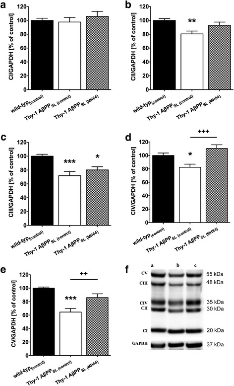
Fig. 4.

Western blot analysis of mitochondrial respiration chain complexes (a CI, b CII, c CIII, d CIV, e CV) in brain homogenate from wild-type(control), Thy-1 AβPPSL (control), and MH84-treated Thy-1 AβPPSL mice. Representative western blots included in lower part of the figure (f). GAPDH was used as loading control. Data represent means ± SEM. N = 11 (six females, five males); one-way ANOVA with Tukey’s multiple comparison post test (***p < 0.001, **p < 0.01, *p < 0.05 against wild-type(control); +++p < 0.001, ++p < 0.01 against Thy-1 AβPPSL (control)). CI complex I (NADH reductase), CII complex II (succinate dehydrogenase), CIII complex III (cytochrome-c reductase), CIV complex IV (cytochrome-c oxidase), CV complex V (F1/F0-ATPase), AβPP beta-amyloid precursor protein
