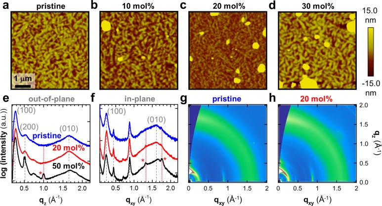
Figure 4.
Atomic force microscopy (AFM) height images of (a) pristine, and N-DMBI-doped p(gNDI-gT2): (b) 10, (c) 20, and (d) 30 mol % N-DMBI. X-ray diffractograms of pristine and doped p(gNDI-gT2) obtained by integration along the (e) out-of-plane (qz) and (f) in-plane (qxy) direction. Scattering from lamellar and π-stacking is indicated with (h00) and (0k0); scattering marked with an asterisk (*) is associated with the neat dopant. 2D grazing-incidence wide-angle X-ray scattering images of (g) pristine p(gNDI-gT2) and (h) the polymer doped with 20 mol % N-DMBI.

