
Fig. 5.
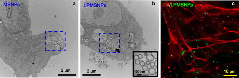
Internalization of LDL peptide decorated PLA-coated MSNPs by RBECs. a TEM image of MSNPs (indicated by blue square) internalized by a RBEC. b TEM image of LDL peptide decorated PMSNPs (indicated by blue square) internalized by a RBEC, scale bar = 2 μm. The inset is an enlarged TEM image verifying the circular structures in side the cell are indeed mesoporous MSNPs. Scale bar = 100 nm. c Fluorescent images showing RBECs (cell membranes were stained with DiI, red fluorescence) cultured with LPMSNPs (green fluorescence). Many green LPMSNPs are seen inside of the RBEC cells. Scale bar = 10 μm