
Fig. 6.
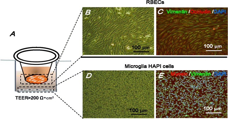
The BBB model. A RBECs are cultured on semi-permeable membranes of Transwell chamber with the HAPI microglia cells grown on the bottom of the well. BBB monolayers with TEER above 200 Ω cm2 were used for transport studies. The same density of RBECs and HAPI cells are also culture in 24-well plates for immunofluorescent staining and imaging. B, D RBECs and microglia HAPI cells were visualized under light microscope respectively. C RBECs were stained with fluorescently labeled antibodies against Vimentin (green) and occludin (red). E HAPI cells were stained with antibodies against Vimentin (green) and Myosin (red). The nucleus is stained by DAPI (blue), scale bar = 100 μm