
Fig. 1.
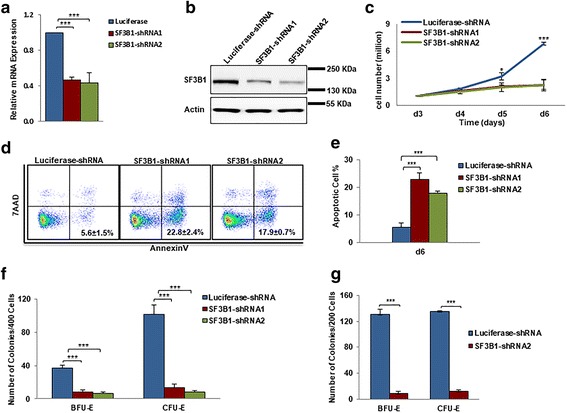
Effects of SF3B1 knockdown on proliferation of erythroid progenitors. a qRT-PCR results showing SF3B1 mRNA expression levels in erythroblasts transduced with lentivirus containing luciferase-shRNA or SF3B1-shRNA, and cultured for 6 days. β-actin was used as internal calculator. Bar plot represents mean ± SD of triplicate samples. b Representative western blotting showing SF3B1 protein levels in erythroblasts transduced with lentivirus containing luciferase-shRNA or SF3B1-shRNA, and cultured for 6 days. c Growth curves of cells transduced with lentivirus containing luciferase-shRNA or SF3B1-shRNA. d Representative flow cytometry profiles of apoptosis as assessed by dual staining of Annexin V and 7AAD at day 6 of culture. e Quantitative analysis of apoptosis from three independent experiments. f Colony-forming ability of cells cultured for 6 days, which contain mixed populations of cells that include BFU-E cells, CFU-E cells, and proerythroblasts. g Colony-forming ability of sorted BFU-E and CFU-E cells using IL-3R, CD34, and CD36 as surface markers [30]. ***P < 0.001