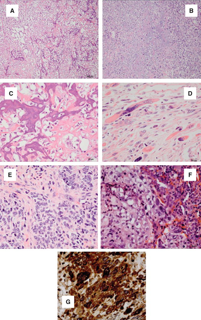
Figure 3. EmCa derived from EmCa stem cells (ECSC). (A, B) Osteosarcomas; (C, D) chondrosarcomas; (E) rhabdosarcoma; (F) epithelial sarcomatous and (G) choriocarcinoma differentiation.
(A) Sarcomatous stromal cells produce osteoid. Tumoral osteoid represented by amorphous fibrillary eosinophilic deposits. Early osteoid deposition forms a lace-like pattern around tumor cells, and advanced osteoid shows evidence of mineralization (darker pink-purple color) (HE, 10×). (B) High magnification shows highly malignant cells with high nuclear-to-cytoplasmic ratio, anaplastic hyperchromatic nuclei or clearing of the chromation and conspicuous, cherry-red nucleoli (HE, 40×). (C) Sarcomatous stromal cells produce chondroid matrix (HE, 10×). (D) Malignant nuclear anaplastic features (HE, 40×). (E) Anaplastic rhabdomyosarcoma showing large strap cells with abundant cytoplasm and striations. The cells are mononuclear or multinucleated. Numerous mitoses are identified (HE, 40×). (F) Epithelial and sarcomatous component blend in this tumor. The malignant epithelial component is composed of cells with high nuclear-to-cytoplasm ratios that show numerous mitoses. Spindle cells with large anaplastic nuclei and prominent nucleoli representing the sarcomatous component are immediately adjacent to the epithelial component (HE, 40×). (G) Malignant polygonal/round cells with single nucleus are reminiscent of the cytotrophoblast. Few multinucleated cells reminiscent of the syncytiotrophoblast are also present. Hemorrhagic background and necrosis areas are present in this tumor (HE, 40×).

