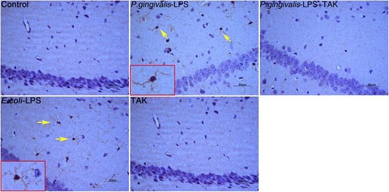
Fig. 6.

Effects of P.gingivalis-LPS on microglia in the hippocampus. Histopathological analysis of brain sections was performed using immunohistochemistry. Microglia were visualized with ionized calcium-binding adaptor molecule 1 (Iba1) (arrows). Activated microglia with irregular protrusions were observed in the P.gingivalis-LPS group (bar = 50 μm)
