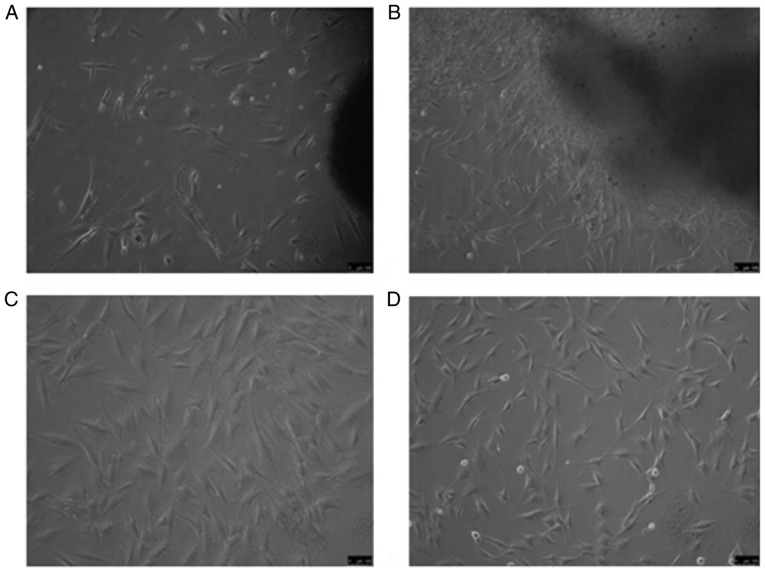
Figure 1.
Fibroblasts derived from hypertrophic scar and normal skin under an inverted phase contrast microscope. (A) Hypertrophic scar tissue adherence after 7 days; (B) normal skin tissue adherence method for 7 days, resulting in long spindle- or star-shaped cells; (C) third generation fibroblasts from hypertro-phic scar source; (D) third generation fibroblasts of normal skin origin (magnification, ×100; scale bar, 100 µm).

