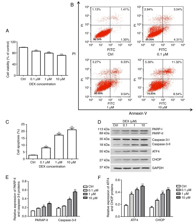
Figure 1.
DEX induces ER stress and apoptosis in MC3T3-E1 cells. (A) The viability and (B and C) apoptosis of MC3T3-E1 cells were affected by DEX in a dose-dependent manner, as determined by a Cell Counting Kit-8 assay and Annexin V-FITC/PI staining, respectively. (D) Western blot analysis of ER stress-associated proteins (ATF4 and CHOP) and apoptosis-associated proteins (PARP and caspase-3). Values are expressed as the mean ± standard deviation (n=3). (E and F) Data from intensities analyses are presented as the relative expression of each protein to GAPDH. *P<0.05 and **P<0.01 vs. Ctrl. Ctrl, control; ATF, activating transcription factor; CHOP, CCAAT/enhancer-binding protein homologous protein; DEX, dexamethasone; ER, endoplasmic reticulum; PARP, poly[ADP ribose] polymerase; PI, propidium iodide; FITC, fluorescein isothiocyanate.

