FIGURE 9.

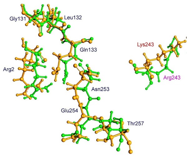
Spatial arrangement of trifluralin-binding amino acids in WT and the Arg-243-Lys mutant α-tubulin isoforms. Residues of WT and mutant isoforms are colored by green and orange, respectively.

Spatial arrangement of trifluralin-binding amino acids in WT and the Arg-243-Lys mutant α-tubulin isoforms. Residues of WT and mutant isoforms are colored by green and orange, respectively.