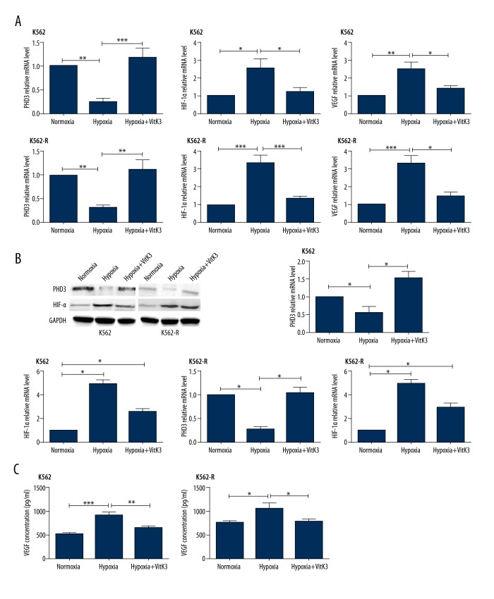
Figure 5.
PHD3, HIF-1α, and VEGF expression levels of both cell lines were evaluated via QPCR, western blot, and ELISA after hypoxia and VitaminK3 stimulation. (A) PHD3, HIF-1α, and VEGF mRNA expression levels of K562 and K562-R cells with or without VitaminK3 stimulation under normoxic or hypoxic conditions. (B) PHD3 and HIF-1α protein immunoactivity of both cell lines on western blots under normoxia, hypoxia, and hypoxia + VitaminK3 treatment. (C) VEGF secretion of both cell lines after hypoxia and/or VitaminK3 stimulation. *** P<0.001; ** P<0.01; *P<0.05.

