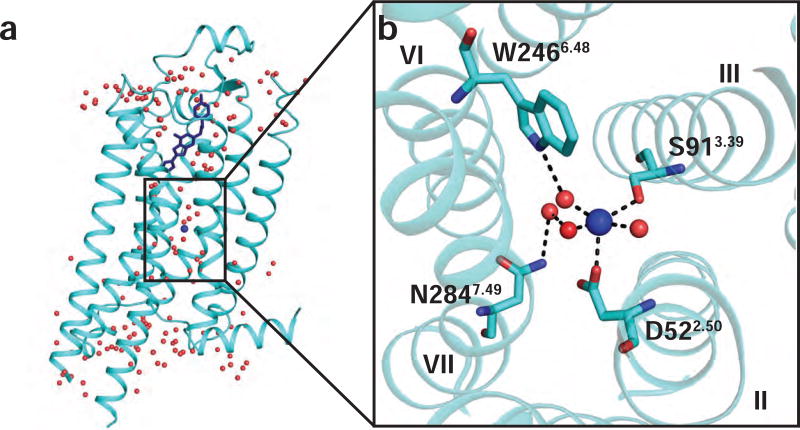
Figure 1. Allosteric sodium binding pocket and sodium coordinating residues in the 1.8 Å A2AAR crystal structure.
(a) Side view of the overall crystal structure of A2AAR in complex with the antagonist ZM241385. A2AAR is shown as cartoon representation and ZM241385 is shown in blue stick representation (PDB ID 4EIY) (Liu et al., 2012). Water molecules are shown as red spheres and sodium is shown as a blue sphere. (b) Expansion of the allosteric sodium pocket. Sodium is shown near the center as a blue sphere and water molecules are shown as red spheres. Residues coordinating with the sodium ion or nearby water molecules are labeled. Dashed lines indicate polar contacts and helices are labeled with Roman numerals.

