
Figure 2. Implanted intracranial electrodes superimposed on 3D reconstruction of the cerebrum.
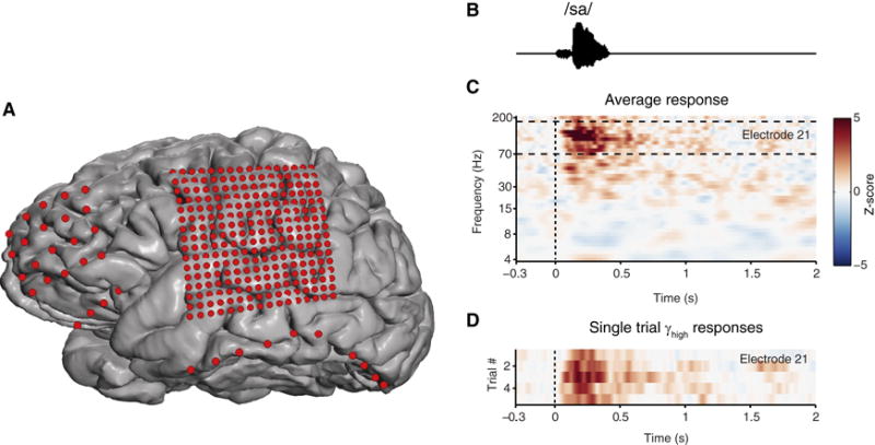
A. Electrode position and relative size in red. Standard 1 cm spaced array over frontal lobe (20 electrode), and ‘high-density’ 4mm array over the lateral cortex covering peri-Sylvian regions (256 electrodes). Subtemporal strip electrodes. Exposed area of electrodes are to scale. B. Speech sound stimulus acoustic waveform. C. Example neural response spectrograms from two neighboring electrodes (z-score) on the superior temporal gyrus. D. Single-trial, high gamma rasters at individual electrodes.