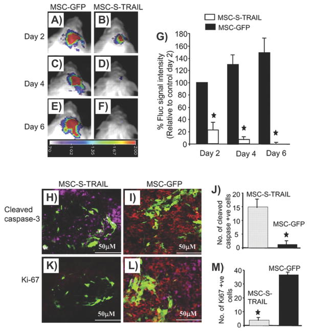
Figure 13.
Mesenchymal stem cells genetically engineered to secrete TRAIL to enhance the treatment of glioma. A–F) Serial in vivo bioluminescence imaging of tumor growth following intracranial implantation of Gli36-EGFRvIII-FD glioma cells mixed with MSCs expressing S-TRAIL (MSC-S-TRAIL; B,D,F) or GFP (MSC-GFP; A,C,E). G) Relative mean bioluminescent signal intensities after quantification of in vivo images. H–M) Photomicrographs show the presence of cleaved caspase-3 (H) and Ki67-positive cells (K) in brain sections from MSC-S-TRAIL-treated and control mice (I,L) 6 days after implantation. Plot shows the number of cleaved caspase-3 (J) and Ki67 (M) cells in MSC-S-TRAIL and MSC-GFP-treated tumors. (Green, MSCs; red, glioma cells; purple, Ki67 or cleaved caspase-3 expression). Reproduced with permission.[265] Copyright 2009, PNAS.

