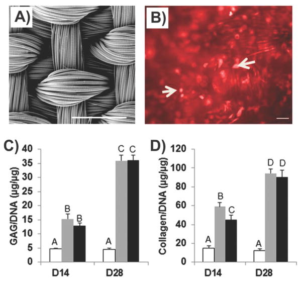
Figure 8.
Engineered stem cells expressing TGF-β3 combined with scaffold for cartilage repair. A) Scanning electron micrograph showing the architecture of the 3D orthogonal woven PCL scaffold. (Scale bar, 500 μm). B) Fluorescence microscopy from iLVT constructs after 28 days in chondrogenic culture. C,D) Quantification of cartilaginous ECM components in the nontransduced (NT), rhTGF-β3 (rhT), and immobilized lentiviral TGF (iLVT) groups. Sulfated glycosaminoglycan content and total collagen content were normalized to DNA content. Bars represent means ± SEM (n = 6). Groups not sharing the same letter or symbol are statistically different (p < 0.05). Reproduced with permission.[185] Copyright 2014, PNAS.

