Figure 1:

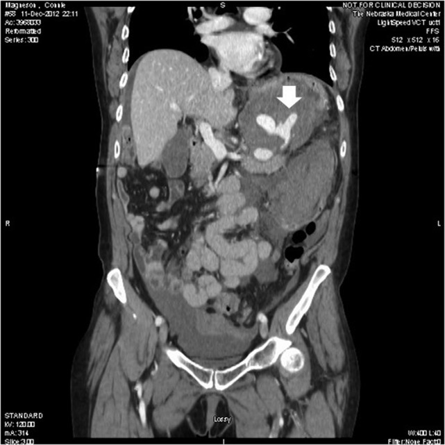
Computed tomography showed three saccular splenic artery aneurysms with a large left upper quadrant hematoma (block white arrow) and blush from distal splenic aneurysm.

Computed tomography showed three saccular splenic artery aneurysms with a large left upper quadrant hematoma (block white arrow) and blush from distal splenic aneurysm.