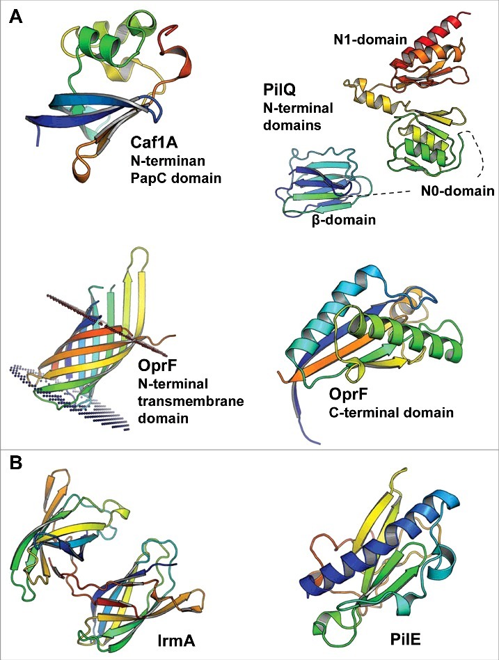
Figure 1.

The 3D structures of bacterial cytokine-binding proteins colored from the N-terminus (blue) to the C-terminus (red). (A) The N-terminal PapC domain of Y. pestis Caf1A (PDB:4BOE), the β, N0 and N1 domains of N. meningitidis PilQ (PDB:4AV2), the N-terminal transmembrane domain (PDB:4RLC), and the C-terminal domain (PDB:5U1H) of P. aeruginosa OprF are located in the outer membrane of gram-negative species. (B) E. coli IrmA (PDB:5EK5) and N. meningitidis PilE (PDB:5JW8) are secreted to and face the extracellular space, respectively. The figures were prepared with PyMol (www.pymol.org).
