
Figure 6.
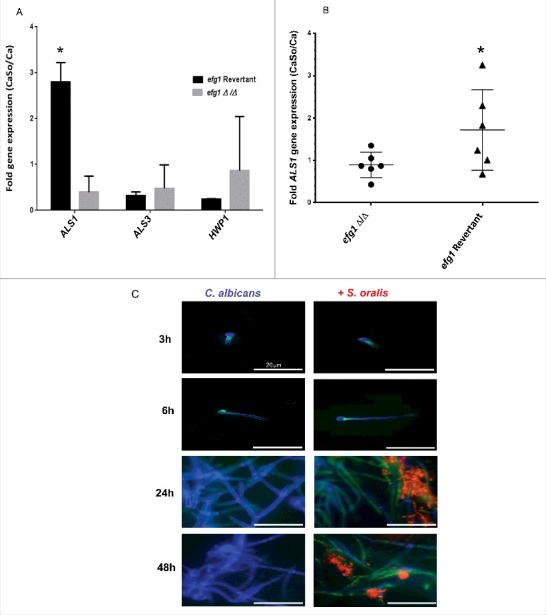
C. albicans ALS1 expression is increased by S. oralis. (A) Biofilms of the efg1 mutant and efg1 revertant were grown with or without S. oralis 34 on the surface of organotypic oral mucosal constructs for 16h. mRNA levels of 3 Candida genes known to play a role in the interactions with oral mucosal epithelium (ALS1, ALS3 and HWP1) were analyzed by RT-qPCR. Results represent fold increase gene expression of each Candida strain with S. oralis over Candida alone. Means ± SD are shown from technical triplicates, in 2 independent experiments. ALS1 was the only gene with expression significantly increased by S. oralis in the efg1 revertant but not in the efg1Δ/Δ strain. *p<0.01 in a comparison between the efg1Δ/Δ and efg1 revertant strains. (B) ALS1 gene mRNA expression levels in tongue tissues of infected mice analyzed by RT-qPCR. Mice were infected with the efg1 mutant (efg1Δ/Δ) and efg1 revertant strain with or without S. oralis 34 for 4 d. Results show ALS1 gene expression levels of the mixed infection group (CaSo) relative to Candida (Ca) infection alone, in 6 mice per group. ALS1 expression was enhanced by S. oralis in the efg1 revertant but not the efg1Δ/Δ infection group. p<0.05 for a comparison between mutant and revertant strains. (C) Als1 protein expression in single (C. albicans) and mixed (C. albicans with S. oralis) biofilms. C. albicans SC5314 was grown on Permanox® plastic chamber slides with or without S. oralis 34 in RPMI 10%FBS, 10% BHI media, for 3h-48 hours. Biofilms were labeled with a monoclonal antibody against Als1, followed by a secondary FITC-conjugated antibody (green). S. oralis (red) was labeled with an Alexa Fluor 568-labeled FISH probe and C. albicans (blue) was stained with Calcofluor White®. A representative of 3 independent experiments is shown. S. oralis increased C. albicans Als1 protein expression on the surface of hyphae after 24–48 h of co-culture. Bars: 50 μm.