
Figure 1.
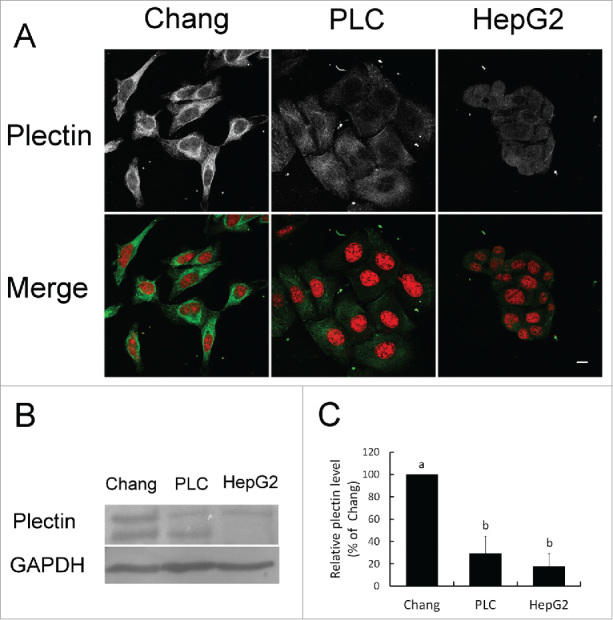
Plectin expression in liver and hepatoma cell lines. (A) Localization of plectin identified by immunofluorescent staining. Red: nucleus. Green: plectin. (B) Western blotting analysis of plectin protein expression in Chang liver cells, PLC/PRF/5, and HepG2 cell lines. (C) Quantification on the level of plectin expression.