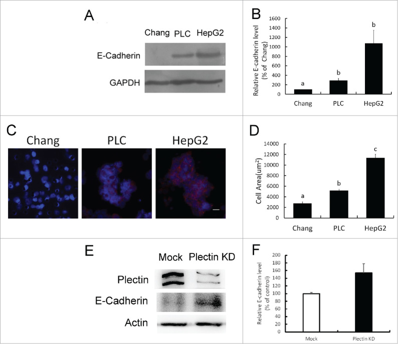
Figure 3.

Collective cell migration associated with E-cadherin expression and plectin deficiency. (A) Western blotting assay of E-cadherin expression in Chang liver cells, PLC/PRF/5, and HepG2 cell lines. (B) Quantification data of E-cadherin expression obtained from Western blotting analysis. HepG2 cells showed highest level of E-cadherin expression compared with Chang and PLC/PRF/5 cells (p < 0.05). (C) Immunofluorescent staining for detecting the distribution of E-cadherin in collective migrated cells. Blue: nucleus. Red: E-cadherin. (D) Quantification of collective cell migration in transwell assay. Cell area appearing more than 3 cells connected with E-cadherin was calculated as collective cell migration. The result of quantification showed significant differences (p < 0.05). (E) Western blotting assay of plectin and E-cadherin expression in mock (Mock) and plectin knockdown-Chang liver cells (Plectin KD), respectively. (F) Quantification data of E-cadherin expression obtained from Western blotting analysis. Plectin knockdown-cells showed higher level of E-cadherin expression compared with mock cells (p < 0.05).
