
Figure 7.
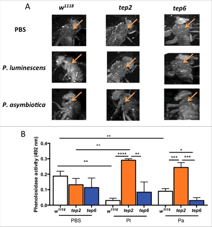
Melanization response and PO activity are elevated in D. melanogaster tep2 mutants upon Photorhabdus infection. (A) Melanization of the wound site in tep2 and tep6 loss-of-function mutant flies and their background control strains (w1118) is shown at 10X magnification 3 h after injection with PBS, P. luminescens or P. asymbiotica bacteria. Arrows indicate the site of injury. (B) PO activity in the hemolymph plasma of tep2, tep6 mutants and control flies (w1118) at 3 hpi with PBS, P. luminescens (Pl) or P. asymbiotica (Pa) (n = 20 flies) as measured by the optical density at 492 nm after incubation with L-Dopa. Values represent the means from 3 biologic replicates and error bars represent standard deviations. Significant differences are indicated with asterisks (*p < 0.05, **p < 0.01, ***p < 0.001, ****p < 0.0001). The means from 3 independent experiments are shown and error bars represent standard deviation.