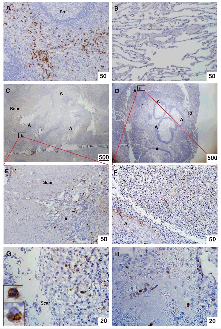
Figure 1.

Immunohistochemical localization of M-ficolin to the aspergilloma. Brown staining indicates presence of M-ficolin. (A) Control immunostaining of monocytes/granulocytes in the spleen. (B) Control alveolar tissue. Overview of elongated A. fumigatus fungal balls surrounded by pulmonary scar tissue in patient 1 (C) and patient 2 (D). The boxes indicate the location of images E-G. (E) Pulmonary scar tissue and A. fumigatus mycelial zone. (F-G) Peripheral zone of aspergilloma (Upper insert: granulocyte. Lower insert: monocyte). (H) Pulmonary blood vessels in scar tissue from a lung with A. fumigatus infection. Fo = follicle. A = A. fumigatus. Scar = scar tissue. The lengths of the bars are in micrometers.
