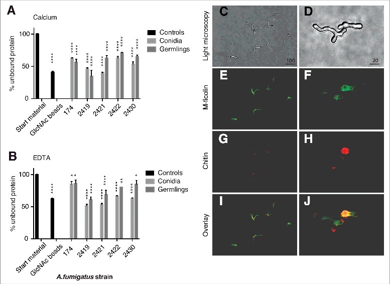
Figure 2.

Characterization of M-ficolin binding to A. fumigatus. The binding between M-ficolin and A. fumigatus strains NRRL 174 (174), SZMC 2419 (2419), SZMC 2421 (2421), SZMC 2422 (2422) and SZMC 2430 (2430) was on conidia (0 hours) and germlings (8 hours) using pull-down assays in the presence of (A) 5 mM Ca2+ or (B) 10 mM EDTA. The data are triplicates from 2 independently performed experiments. Data shown are mean ± SEM. Significance was determined using 2-way ANOVA with Holm-Sidak's multiple comparison test, *p < 0.05, **p < 0.01, ****p < 0.001. (C) Light microscopy of growing hyphae, original magnification 100×. (D) Light microscopy of growing hyphae, original magnification 400×. (E-F) Localization of regions recognized by M-ficolin (green) and (G-H) localization of chitin (WGA, red). (I-J) Overlay images. The lengths of the bars are in micrometers.
