Figure 2.
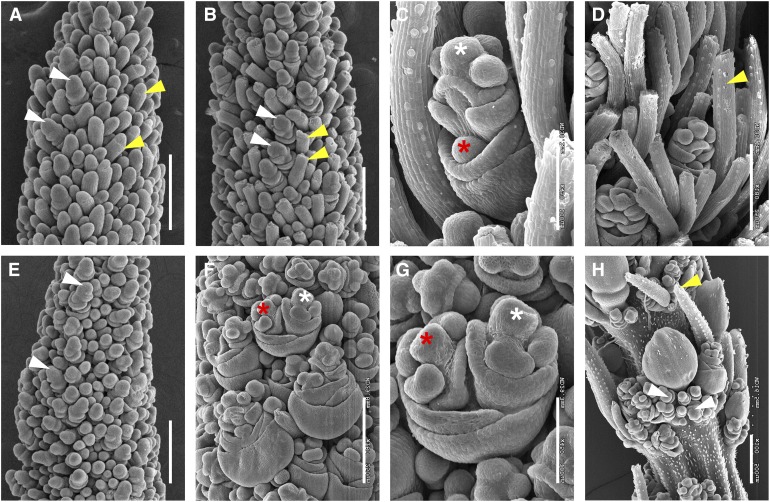
Morphological Characterization of Inflorescence Development in the bsl1-1 Mutant by Scanning Electron Microscopy Analysis.
(A) and (B) Bristle development was first apparent at 17 DAS in A10.1 wild-type inflorescence primordia (A) and bristle differentiation was obvious by 18 DAS where meristem tips appeared to break off at indentations marked by yellow arrows (B). Differentiating spikelets are marked by white arrows. Bars = 250 μm.
(C) and (D) At 20 DAS, bristles were well developed in the wild-type inflorescences, and within spikelets, an upper floret (white asterisk) developed and a lower floret was visible as a meristematic bulge prior to abortion (red asterisk; [C]). A mature bristle is marked with a yellow arrow (D). Bars = 100 μm in (C) and 250 μm in (D).
(E) In the bsl1-1 mutant inflorescence primordium, bristles were not initiated by 18 DAS, but spikelets appeared to develop normally (white arrows). Bar = 250 μm.
(F) to (H) At 20 DAS, bristle development was highly reduced in the bsl1-1 mutant and upper (white asterisk) and lower (red asterisk) florets were both developed (G). Precocious development of numerous rudimentary spikelets (white arrows) was observed at the base of main spikelets (H). Yellow arrow indicates one of few differentiated bristles formed in the mutant. Bars = 250 μm in (F) and (H) and 100 μm in (G).

