
Figure 10. Echocardiogram from child with severe mitral regurgitation.
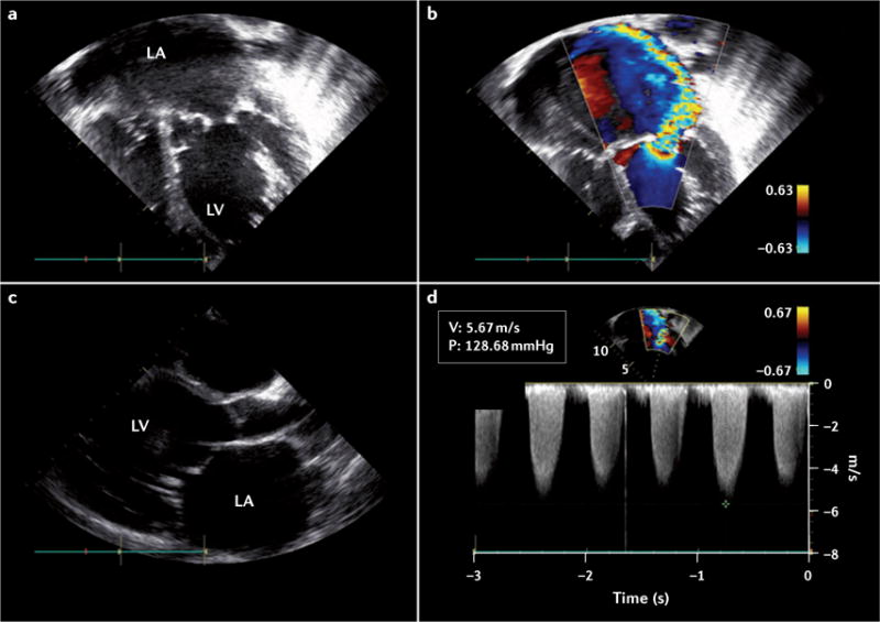
This echocardiogram was produced as part of an echocardiography screening programme. a | Apical four-chamber view in black and white Doppler. b | Apical four-chamber view in colour Doppler. The colour jet extends to the back of the left atrium. c | A parasternal long-axis view. The mitral valve is thickened with excessive leaflet tip motion and lack of coaptation. The left atrium is severely dilated and the left ventricle is moderately dilated. d | Pan-systolic spectral Doppler of mitral regurgitation. LA, left atrium; LV, left ventricle; P, pressure; V, volume.