
Figure 11. Echocardiogram from child with severe mitral stenosis.
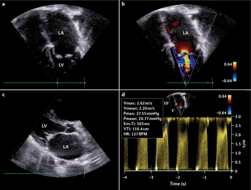
This echocardiogram was produced as part of an echocardiography screening programme. a | Apical four-chamber view in black and white Doppler. b | Apical four-chamber view in colour Doppler. The colour jet reveals turbulence in diastole. c | A parasternal long-axis view. The mitral valve is thickened with limited motion and the chordae are thickened and fused. The left atrium is severely dilated. d | Pan-diastolic spectral Doppler of with severe mitral stenosis mean gradient (mean 21 mmHg). Env. Ti, envelope time; HR, heart rate; LA, left atrium; LV, left ventricle; Pmax, maximum pressure gradient; Pmean, mean pressure gradient; Vmax, maximum velocity; VTI, velocity time integral.