
Figure 4. Molecular and cellular basis of Sydenham’s chorea.
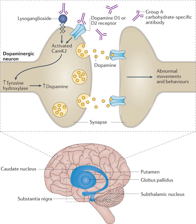
In Sydenham’s chorea, neurons in the basal ganglia are attacked by antibodies against the group A carbohydrate of Streptococcus spp. that react with the surface of the neuron. This reaction activates signalling through calcium/calmodulin-dependent protein kinase type II (CAMK2), which involves an increase in tyrosine hydroxylase in dopaminergic neurons. Receptors, such as the D1 and D2 dopamine receptors, and lysoganglioside might be autoantibody targets on the neuronal cell. This targeting could lead to altered cell signalling and increased levels of dopamine, in turn leading to abnormal movements and behaviours.