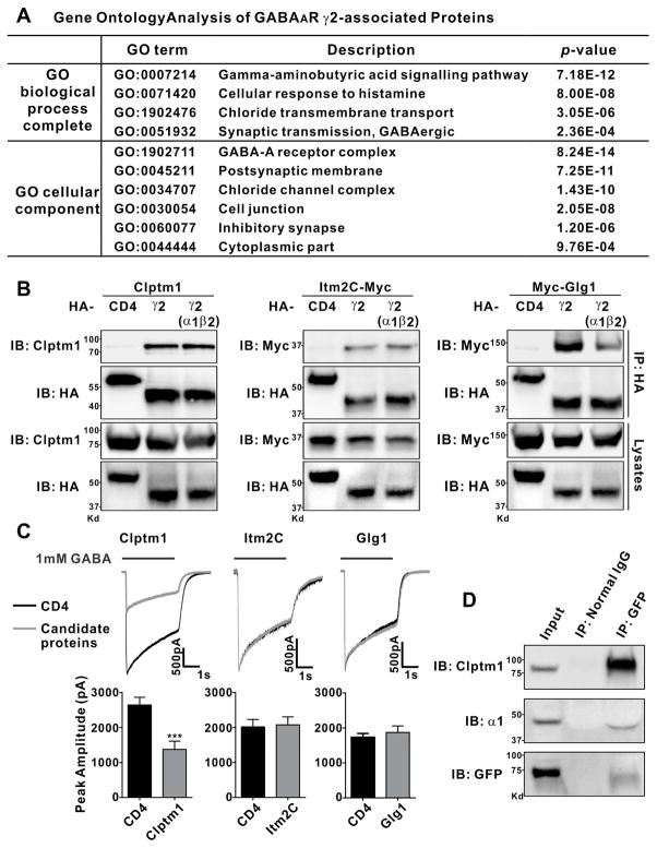
Figure 2. Clptm1, Itm2C, and Glg1 Associate with GABAAR γ2.
(A) For gene ontology analysis, the set of HFY-GABAARγ2-associated proteins listed in Table S1 was compared with the entire mouse database to identify pathways and compartments in which these proteins are significantly enriched using the statistical overrepresentation test (PANTHER with Bonferroni correction for multiple testing). Terms with p value <0.001 are listed here.
(B) HEK293 cells were transfected with Clptm1, Itm2C-Myc, or Myc-Glg1 and HA-tagged GABAAR γ2 alone, GABAAR γ2 with non-tagged α1 and β2, or HA-CD4 as a negative control. Clptm1, Itm2C, and Glg1 were specifically co-immunoprecipitated with GABAAR γ2 but not CD4.
(C) HEK293 cells which stably express GABAARα1/β2/γ2 were transfected with Clptm1, Itm2C, Glg1, or HA-CD4, each together with GFP to detect the transfected cells for recording. GABAAR mediated currents were induced by fast application/removal of GABA (1 mM). Clptm1 significantly reduced GABAAR mediated currents. Clptm1: n=18 cells from 3 independent experiments, Itm2C: n=17 cells from 2 independent experiments, and Glg1: n=21 cells from 2 independent experiments. *** p<0.001, t-test.
(D) Clptm1 was co-immunoprecipitated with GABAAR γ2 and α1 in whole brain homogenate from transgenic HFY-GABAARγ2 mice.
Results are expressed as mean ± SEM.

