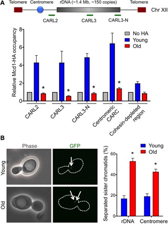
Fig. 2. Cohesion is lost during replicative aging.

(A) ChIP analysis of hemagglutinin (HA)–tagged Mcd1 in young and old cells. An untagged strain was used as a negative control. The average and SEM of four replicates are plotted. *P < 0.05, as determined by Student’s t test. (B) Loss of cohesion at rDNA and centromeric regions with aging. Representative images are shown on the left. The average and SEM of three replicates are plotted. The arrows indicate one spot or two spots of GFP lac repressor. *P < 0.05, as determined by Student’s t test.
