
Figure 1.
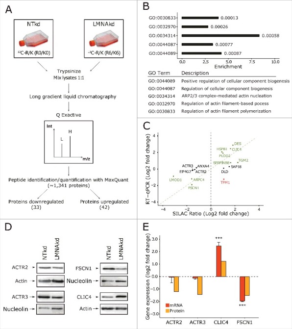
SILAC based quantitative proteomics of human dermal fibroblasts after sustained LMNAkd indicate that depletion of lamin A/C influences the levels of proteins involved in actin cytoskeleton organization. (A) Schematic overview of the SILAC experiment (see materials and methods for details). L = light medium, H = heavy medium, Int = intensity, m/z = mass/charge ratio. (B) Proteins that showed significant differences (p < 0.05) in the SILAC dataset were analyzed with Gorilla, yielding a subset of significantly affected pathways (p-value for the respective GO term depicted next to the bars). (C) Scatterplot between mRNA level, measured by RT-qPCR relative to non-targeting control (NTkd), and SILAC ratio. Genes in green text color showed correlation, genes in black text color showed no correlation, and genes in red text color showed inverse correlation. (D) Western blot analysis of ACTR2, ACTR3, FSCN1 and CLIC4. Nucleolin or actin was used as a loading control. (E) Gene expression levels of ACTR2, ACTR3, FSCN1 and CLIC4 measured by real-time qPCR relative to NTkd (in orange) and protein levels of ACTR2, ACTR3, FSCN1 and CLIC4 semi-quantified with western blot analysis (in yellow). Error bars indicate the standard deviation on the mean value of 3 biological replicates; significance was calculated with a Student's t-test; *** = p-value < 0.001.