
Figure 4.
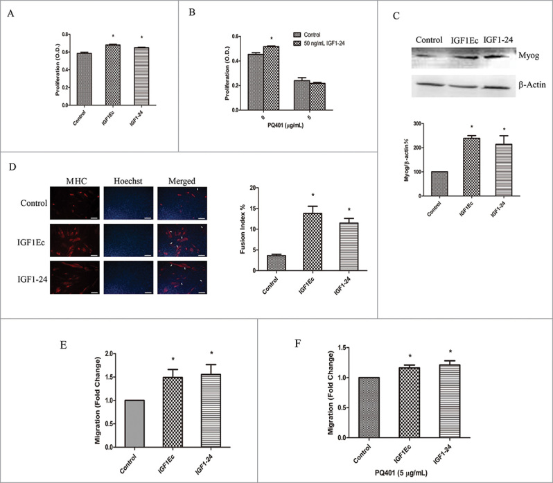
(A)recombinant protein IGF1–24 has similar function with IGF1Ec. (A) C2C12 cells were treated with IGF1Ec and IGF1–24 (50 ng/mL) for 48 h, and proliferation efficiency was measured via CCK-8. (B) After pretreated with PQ401 (5 μg/mL), C2C12 cells were treated and analyzed as in (A). (C) C2C12 cells were treated with IGF1Ec and IGF1–24 (50 ng/mL) for 5 d in DM. The expression of Myog was detected by western blot. (D) C2C12 cells were treated with IGF1Ec and IGF1–24 (50 ng/mL) for 5 d in DM. Skeletal muscle MHC (terminally differentiated state marker) was detected via immunofluorescence (red). Nuclei were visualized using DNA Hoechst staining (blue). Fusion index was defined as the percentage of nuclei belonging to MHC positive cells with 3 or more nuclei. (E) C2C12 cells were seeded into the upper chambers in serum-free media, and 50 ng/mL IGF1Ec and IGF1–24 were placed in the bottom chambers. After 6 h migration, migrated cells were stained with 0.1% crystal violet, imaged and counted. (F) After pretreated with PQ401 (5 μg/mL), C2C12 cells were treated and analyzed as in (E). Columns, mean of at least 3 independent experiments; Error bars, SEM. *, p < 0.05. Bars, 100 μm. Arrows indicate multi-nucleated myoblast fusion.