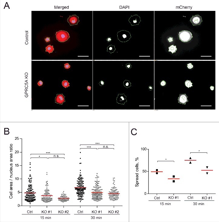
Figure 2.

GPRC5A affects cell spreading. (A) GPRC5A-KO MDA-MB-231 cells demonstrate slower spreading on Collagen I-coated (0.1 mg/mL) surface compared with isogenic control. Representative images show nuclear (DAPI) and the whole cell area (mCherry) 30 min after plating. Scale bars correspond to 100 µm. (B) At least 60 cells were quantified for each sample and plotted as the nucleus / whole cell area ratios 15 or 30 min after plating for 2 independent experiments with 2 technical replicas in each (N = 2, n = 2). Red lines represent mean values. (C) The number of flattened cells (for which the total/nucleus area ratio was greater than 3) was smaller for GPRC5A knock-out cells (KO) compared with control (Ctrl). Statistical significance was evaluated using ANOVA with Tukey post-hoc test: *, p < 0 .05; **, p < 0 .01; ***, p < 0 .001.
