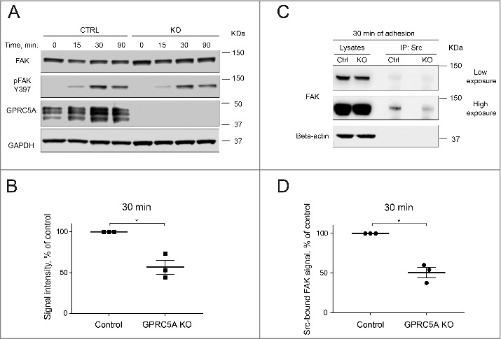
Figure 4.

Knock-out of GPRC5A results in impaired activation of FAK in adhering cells. (A) A representative Western blot demonstrating FAK phosphorylation dynamics in MDA-MB-231 cells at different time points after plating on Collagen I (0.1 mg/mL). (B) Quantification of the Western blot shown in (A) for 30 min after plating. Phosphorylated FAK signal was normalized to the loading control (GAPDH) and total FAK signal. Data represent the mean ± SEM, N = 3. Statistical significance was evaluated using one-way ANOVA test. *, p < 0 .05. (C) Representative Western blot demonstrating the amount of FAK co-immunoprecipitated with Src in control and GPRC5A knock-out MDA-MB-231 cells 30 minutes after plating on Collagen I (0.1 mg/mL). (D) Quantification of the Western blot shown in (C). Src-co-immunoprecipitated FAK signal was normalized to loading control and total FAK signal of the input samples. Data represent means ± SEM, N = 3. Statistical significance was evaluated using one-way ANOVA test. *, p < 0 .05.
